

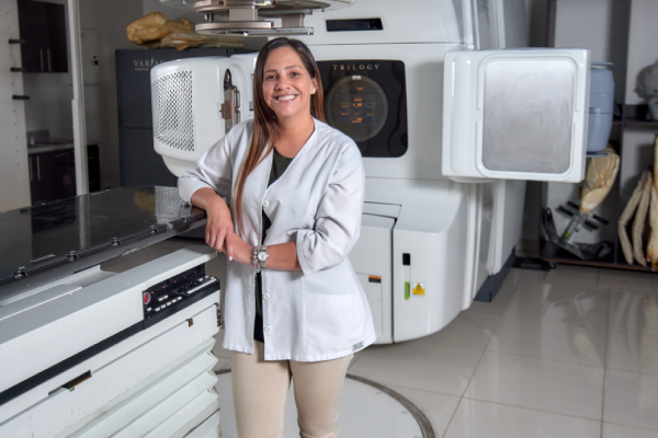
Specialist with extensive experience in generalized radiation oncology treatments. Extensive knowledge in radiosurgery applied to head and neck tumors and prostate cancer. Extensive experience in novel schemes in the treatment of breast cancer.
UCIMED University of Medical Sciences | From 2010
Postgraduate Systems, University of Costa Rica | Since 2015
Mexico Hospital | 2011 - 2015
Mexico Hospital | 2015 - Currently

Specialist with extensive experience in radiosurgery treatments. He has knowledge of novel approaches and techniques for intracranial and CNS injuries. Dr. Soto has specialized in comprehensive radiation oncology with an emphasis on short and precise treatments.

UCIMED University of Medical Sciences | Since 1998
University of Costa Rica UCR. | Since 2007
Central American Institute of Public Administration ICAP | Since 2017
University of Los Andes, Chile. IAEA-FALP | Since 2018
San Juan de Dios Hospital | 2011 - Current
Radiotherapy Service San Juan de Dios Hospital | 2015 - current
Calderón Guardia Hospital, Mexico Hospital and San Juan de Dios Hospital | 2007 - 2011
National Insurance Institute (INS)
2005 - 2007
CARIBBEAN PRESMED

Dr. Gallegos has extensive experience in radiation treatments, both radiotherapy and radiosurgery. He has experience in the management of multiple and localized injuries with novel approaches.

San Jose, Costa Rica | Since 2019
San Jose, Costa Rica | Since 2018
San Jose, Costa Rica | Since 2017
Argonne, Illinois, USA | Since 2016
San Jose, Costa Rica | Since 2012
University of Costa Rica | Since 2003
University of Costa Rica | Since 2003
Mexico City, DF | Since 2003
University of Costa Rica | 2009 - Current
San Juan de Dios Hospital. Hospital and Mexico | 2003 - Current
Mexico Hospital | 1999 - 2003
United Supermarkets
1998 - 1999
Guanacaste, Ministry of Health | 1997 - 1998

Extensive experience in radiation oncology treatments for gastrointestinal cancer. He also has extensive knowledge in innovative radiosurgery and body radiotherapy schemes.
University of Costa Rica | Since 2008
University of Costa Rica | Since 2015
Arturo López Pérez Foundation -University of the Andes Santiago de Chile | Since 2018
Mercedes Chacón P Clinic | 2009
El Rosario School |
Oscar Lambret Institute, Lille - France | 2014
2017 - 2018
Mexico Hospital | 2018 - Currently


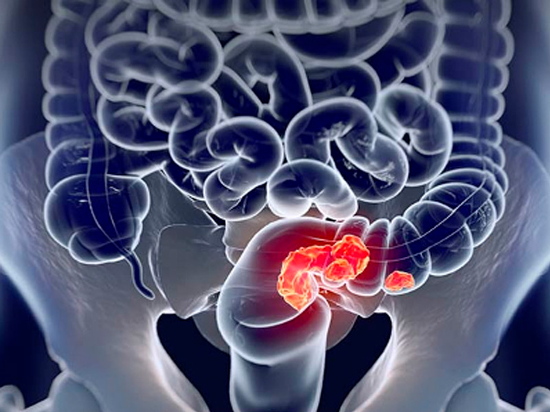
He rectal cancer It is defined as the tumor located between the anal margin and 15 cm proximally. In rectal cancer, accurate preoperative staging allows patients to be correctly classified for the various existing therapies, as well as to select the best surgical treatment.
FAMILY BACKGROUND: Colorectal cancer in a first-degree relative.
PERSONAL HISTORY: From colorectal adenomas, colorectal cancer or ovarian cancer.
Hereditary Conditions: Such as familial adenomatous polyposis (FAP) and Lynch syndrome (hereditary nonpolyposis colon cancer [HNPCC])
EXCESSIVE ALCOHOL CONSUMPTION
TOBACCO
AFRICAN AMERICAN RACE OR ETHNICITY
OBESITY
The symptoms of rectal cancer are similar to those of colon cancer and will depend on the time of evolution and its location (distance from the anal margin, location on the anterior wall, circumferential, etc.), including the following:
SYMPTOMS: Rectal bleeding, Change in bowel habit, Abdominal pain, Intestinal obstruction, Change in appetite, Weight loss, Weakness.
With the exception of symptoms of obstruction, these symptoms do not always correlate with the stage of the disease or signify a particular diagnosis.
DIAGNOSIS: The way to make the diagnosis is within the reach of every doctor and only consists of a digital rectal examination (DRE) in which an irregular tumor mass that is generally hard and usually fixed will be palpated. It is also believed that currently not only the digital rectal examination should be considered in the diagnosis but also the low endoscopic study (colonoscopy) in all patients over 40 years of age who consult for hematochezia (elimination of visible blood from the rectum). The finding of neoplastic lesions in the endoscopic examination will be accompanied by the anatomopathological study of biopsies which should confirm the diagnosis of adenocarcinoma.
In patients with CR, staging is essential not only to estimate the prognosis but also to define the different therapeutic alternatives. Let us remember that the surgeon must decide whether the patient should undergo preoperative chemoradiotherapy, decide the surgical technique (anterior resection with or without preservation of the sphincter apparatus, local trans-anal resection) and discuss in detail any possible sequelae. For all these decisions, it is essential to carry out an optimal study and preoperative staging. This staging will undergo changes in patients undergoing preoperative chemoradiotherapy since as a result of its effectiveness a marked reduction in tumor mass (wall and lymph node) will be obtained in more than 70% of patients and even a complete tumor response in around a 10 to 20% of them.
Below are the details of some of these treatments:
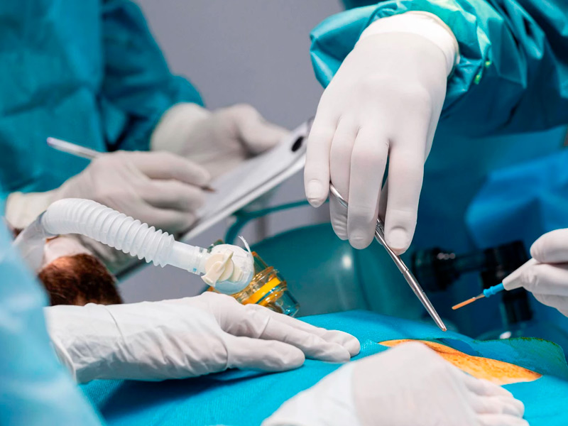
SURGICAL TREATMENT: Surgery is the fundamental basis for the successful treatment of colorectal carcinoma. Its objective is the removal of the primary tumor and any loco-regional extension that may have occurred, without causing tumor spread and with the best quality of life for the patient.
CHEMOTHERAPY TREATMENT: When choosing the chemotherapy regimen to be administered, the activity and tolerance of the chemotherapy regimen and a series of factors that depend on the patient (will, general condition, comorbidity, etc.) are taken into account. It increases survival and can be given palliatively in very advanced cancers.
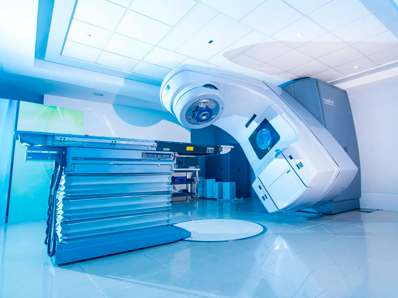
RADIOTHERAPY TREATMENT: The use of radiotherapy as part of the treatment of rectal malignancies is increasingly widespread. In locally advanced primary rectal cancer, several studies have demonstrated its effectiveness, with a decrease in local recurrence and an increase in disease-free survival, both in its preoperative and postoperative administration. Emerging developments such as intensity-modulated radiation therapy (IMRT), image-guided radiation therapy (IGRT), and stereotactic radiation therapy (SBRT) are being evaluated. These techniques offer the possibility of providing greater radiotherapy without involving normal or healthy tissue.
Currently the 21st Century Radiotherapy Clinic has the technology and experience to treat this type of diseases, guaranteeing the protection of surrounding organs and tissues, thus providing a higher quality of life for the patient.
Before and/or after surgery to help prevent the cancer from coming back. In this case, it is often given together with chemotherapy. Many doctors now favor giving radiation therapy before surgery because it can make it easier to remove the cancerous tumor.
With or without chemotherapy to help control rectal cancers in people who are not healthy enough to have surgery or to relieve symptoms in people with advanced cancer that is causing intestinal blockage, bleeding, or pain.
To re-treat tumors that have returned in the pelvic region after undergoing radiotherapy.
To help treat cancer that has spread to other areas (Metastases), such as bones, liver, brain.

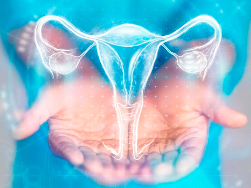
He cervical cancer It begins when healthy cells on its surface begin to divide uncontrollably. These changes cause anomalies, not necessarily cancerous. These are the first steps that can lead to the formation of cancer.
Cervical cancer, or cervix, is among the most frequent in the female population worldwide, and according to statistics from the World Cancer Report 2014, it has fourth place in incidence and mortality. It is estimated that in 2018 some 570,000 new cases, representing 6.6% of cancers in women and a total of 311,365 deaths were recorded, with the populations of less developed regions, included in this group Central America and the Caribbean, being the most affected.
Its incidence among women aged 20-54 years, in Latin America, is 1: 1000 women per year. Costa Rica has an incidence of 20.2/1 00,000 women, which places us among the intermediate risk countries. In Costa Rica, in 2014, cervical cancer ranked third in incidence, being most common in the provinces of San José, Alajuela and Heredia, and fourth in mortality in 2015, with Cartago and Alajuela being the most affected provinces. according to the National Tumor Registry (RNT) and the National Institute of Statistics and Censuses (INEC).

As with other chronic diseases, cervical cancer incidence and mortality rates increase with age, yet the greatest absolute burden of cervical cancer falls on middle-aged women. Cervical cancer represents one of the few common cancers for which a specific causative agent has been identified.
Chronic HPV infection is the underlying cause in more than 99% of cervical cancer cases. In addition to HPV-16 and HPV-18, recent international studies have expanded the list of oncogenic viruses to include types 31, 33, 35, 45, 51, 52, 58 and 59. Globally, there have been the prevalence of HPV in 99.7 percent of cervical carcinomas, and oncogenic types 16 and 18 are those most frequently detected. All those related to the acquisition of the infection are considered risk factors.
There are other cancers related to chronic HPV infection such as cancer of the vulva (46%), vagina (70%), anal (88%), penis (50%) and oropharynx (26-50% depending on the geographic region).
HPV is the most common sexually transmitted infection and the majority of the sexually active population comes into contact with the virus throughout their lives. The infection is asymptomatic so many of those infected are unaware of it and therefore can transmit it. Up to 90% of HPV infections are cleared without treatment during the first two years and only those that become chronic can give rise to precancerous lesions that progress to invasive cancer.
Environmental agents such as tobacco smoke, hormonal contraceptives, diet, and various infectious microorganisms have been evaluated as possible cofactors of HPV in cervical carcinogenesis. Well-controlled epidemiological studies have shown that patients with HPV-positive cervical cancer are twice as likely to have been smokers as HPV-positive controls. Additionally, some recent studies in HPV-positive women have indicated that the risk of cervical cancer is higher in women who have used oral contraceptives for more than 10 years.
In the earliest stages the disease is frequently asymptomatic. The earliest symptoms include:
SYMPTOMS: Abnormal genital bleeding (irregular/intermittent), Bleeding after sexual intercourse (coitorrhagia) or during gynecological examination, Smelly discharge, is very nonspecific, but may be indicative of vaginitis or cervicitis, Pelvic or lower back pain, Discomfort when urinating (dysuria) or Rectal tenesmus, Gynecological bleeding after menopause, Pain during sexual relations (dyspareunia).
DIAGNOSIS: Cytology using the Papanicolaou technique has a low sensitivity for the diagnosis of high-grade lesions, but it compensates with a high specificity. The combination of molecular detection of human papillomavirus (HPV) and cytology reaches a sensitivity to detect these lesions of up to 96%. In a vaccination scenario like the one being implemented for this pathology.
The Pap test has reduced the incidence rate of cervical cancer by 60%-90% and the mortality rate by 90%. More recently, an HPV DNA detection test has been introduced that has greater sensitivity for high-grade CIN than the Pap test and has been shown to provide greater protection against invasive cancer compared to the Pap smear. with the latter.
Physical and gynecological examination: A visual examination of the cervix is performed with a speculum. Those lesions visible with this technique include ulcerations, exophytic tumors in the ectocervix, and infiltration of the endocervix.
Cervical cytology (Pap smear): It is the main population screening method. Identify abnormal cells. Low sensitivity but high specificity.
HPV Test: Test with high sensitivity and specificity.
Colposcopy: It is performed when any of the screening tests are positive or if there is clinical suspicion. It allows us to see the morphology of suspicious lesions in greater detail and take biopsies at the same time.
Cervical biopsy: It consists of taking a small fragment of the suspicious lesion to corroborate the suspected diagnosis.
Image tests:
– Chest x-ray.
– Cystoscopy and/or rectosigmoidoscopy: It consists of performing a direct visualization of the bladder and rectum respectively in case of suspicion of infiltration by the tumor.
– Urography: allows evaluation of the urinary tract in the event of suspicion of locally advanced disease.
– Transvaginal Ultrasound.
– Computed Tomography (CT): It can be used instead of chest x-ray and urography for staging and is also useful for assessing lymph node involvement.
– Magnetic resonance: very useful test for local staging of the disease by determining tumor size, invasion of adjacent tissues and lymph node involvement.
Positron emission tomography (PET): This nuclear medicine imaging test uses a small amount of radioactive material to help determine how much cervical cancer has spread.

Treatment depends on the diagnosis, the size, location and stage of the tumor, as well as your general health and physical condition. Depending on the extent of the cancer and risk assessment, treatment may consist of one or more therapies:
SURGERY: The conventional schedule for delivering radiation to the whole breast is 5 days a week (Monday through Friday) for 6 to 7 weeks. Another option is hypofractionated radiation therapy in which radiation is also given to the entire breast, but in higher daily doses (Monday through Friday) using fewer treatments (usually for only 3 to 4 weeks).
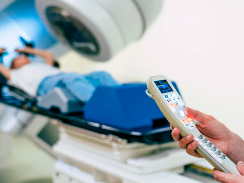
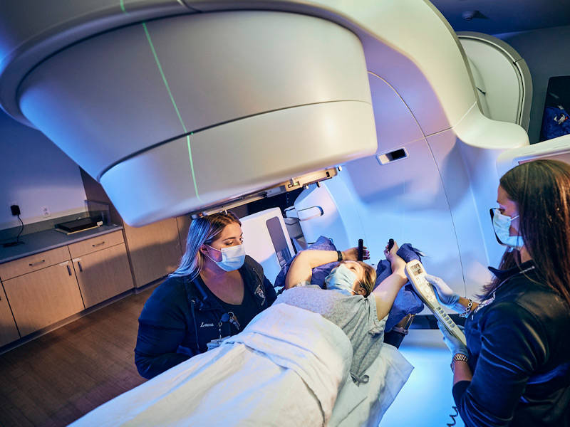
RADIOTHERAPY: Radiation therapy may be given after surgery or instead of surgery, and is the treatment of choice during any stage of the disease, except for the earliest stages. It is also used to treat some patients with locally advanced cervical cancer and can also be used to treat some symptoms of metastatic disease, as neoadjuvant or adjuvant therapy depending on the case. It is a treatment that is administered on an outpatient basis 5 days a week, for approximately 5 weeks. Administration of the radiotherapy treatment lasts a few minutes, and is not painful.
Currently, there are very sophisticated advanced treatment techniques with external radiotherapy that allow high doses of radiation to be administered to the treatment area, minimizing the radiation dose to the healthy organs surrounding the tumor and demonstrating that the overall survival and the disease-free period In this type of pathology, the application of these techniques is favored.
Currently the 21st Century Radiotherapy Clinic It has these technologies and the experience to treat this type of pathologies, guaranteeing the protection of surrounding organs and tissues, with a reduction in toxicities at the level of the urinary tract and the gastrointestinal tract, thus providing a greater quality of life for the patient. These techniques are:
* IMRT (intensity modulated radiotherapy): Allows the RT dose and intensity to be varied during therapy. It is a technology through a computerized system that allows precise radiation doses to be delivered to a tumor or specific areas within a tumor and reduce doses to healthy tissues.
* VMAT (Volumetric Arcotherapy): This technique uses a computer-controlled machine that moves around the patient as it emits radiation and significantly shortens irradiation times, contributing to further increasing the safety of the treatment and improving patient comfort.
* IGRT: Allows the delivery of higher doses of radiotherapy by utilizing advanced imaging techniques such as magnetic resonance imaging with spectroscopy or an integrated imaging scanner. This advancement allows the doctor to take photographs of the area just before administering radiation to make minor adjustments to the direction of the rays, helping to deliver the radiation even more precisely.
CHEMOTHERAPY: The use of radiotherapy as part of the treatment of rectal malignancies is increasingly widespread. In locally advanced primary rectal cancer, several studies have demonstrated its effectiveness, with a decrease in local recurrence and an increase in disease-free survival, both in its preoperative and postoperative administration. Emerging developments such as intensity-modulated radiation therapy (IMRT), image-guided radiation therapy (IGRT), and stereotactic radiation therapy (SBRT) are being evaluated. These techniques offer the possibility of providing greater radiotherapy without involving normal or healthy tissue.

He gastric cancer It is a general term for any malignant tumor that arises from the cells of any of the layers of the stomach. It is the most common neoplasm of the digestive tract worldwide, being the third cause of death from cancer worldwide in both sexes. The term gastric cancer refers to adenocarcinomas of the stomach, the most common histological type, which represent a 95% of malignant tumors of this organ. Except in Japan, carcinoma of the stomach is generally in an advanced stage of development at the time of diagnosis, with infiltration beyond the submucosa and invasion of the gastric wall.

NUTRITIONAL FACTORS: Diets rich in salt and smoked foods typical of Japan, Korea and China, low in fresh fruits and vegetables, and high concentrations of nitrates in foods favor the risk.
ENVIRONMENTAL FACTORS: Poor food preparation, lack of refrigeration and poor water that may have high concentrations of nitrates or Helicobacter pylori increase the risk.
TOBACCO: Tobacco increases the risk of developing many cancers, including stomach cancer.
PREDISPOSING DISEASES OR CONDITIONS: Diseases or predisposing conditions:
Previous gastric surgery: It takes years for cancer to appear on the residual stomach (gastric stump). In general, this period is usually longer than 10 -15 years.
Chronic atrophic gastritis: It can degenerate until it becomes cancer.
Pernicious anemia: It is a special type of anemia, which increases the risk by about 20 times.
Gastric polyps: The risk of cancer developing on a polyp depends, among other factors, on the size of the polyp and its histology. In general, the larger the size, the greater the risk of malignancy.
H. Pylori infection: H. pylori is a bacteria that can be found in the stomach and cause ulcers and chronic gastritis. Worldwide it is the most important risk factor for gastric cancer. However, despite the increased risk of gastric cancer, most people with this infection will NOT develop it.
Gastroesophageal reflux: Increases the risk of cancer of the gastroesophageal junction.
Family Factors
The diagnosis of gastric cancer is based on clinical history, physical examination, blood tests, imaging tests such as CT, upper digestive endoscopy (gastroscopy) and biopsy.
CLINIC
ASYMPTOMATIC: Gastric cancer may not produce symptoms until advanced stages.
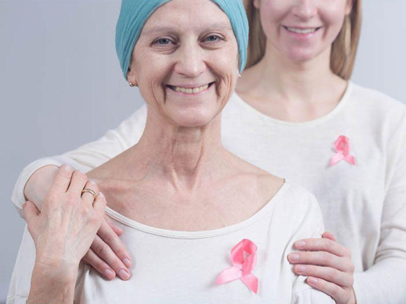
Recent nipple inversion scaly, peeling, crusting, and peeling of the pigmented area of skin surrounding the nipple (areola) or breast skin, Redness or small holes in the skin over your breast , like the peel of an orange.
SYMPTOMS: Symptoms (what the patient notices) are usually vague and nonspecific. The most common are indigestion, weight loss, upper abdominal pain, changes in bowel frequency, loss of appetite, and gastrointestinal bleeding. Bleeding can be of various types and cause anemia. Nausea and vomiting, a feeling of early fullness (feeling of being full after eating little) due to lack of distention of the gastric wall, ascites (accumulation of fluid in the abdomen), fatigue, etc. may also be noted.
Endoscopic mucosal resection (EMR): This REM technique consists of removing the tumor using gastroscopy and is reserved for cancers that are in early stages.
Below are the details of some of these treatments:
SURGERY: Gastrectomy is the standard surgical technique to resect the primary tumor.
RADIOTHERAPY: It is treatment with ionizing radiation with technologies such as intensity modulated radiotherapy (IMRT). These treatments use special computers and techniques to focus radiation on the cancer and limit damage to adjacent normal tissues. The patient lies on the table and has to remain still for the minutes that the radiotherapy administration lasts. Radiotherapy is NOT a painful treatment.
Currently, the Siglo XXI Radiotherapy Clinic has the technology and experience to treat this type of disease, guaranteeing the protection of surrounding organs and tissues, thus providing a higher quality of life for the patient.
To treat gastric cancer, radiotherapy can be used in different ways:
After surgery called adjuvant radiation therapy can be used to destroy small remnants that cannot be removed with surgery. Radiotherapy can be accompanied by chemotherapy to prevent or postpone recurrence and increase its effectiveness.
Less frequently, it can be administered before surgery in a neoadjuvant manner with chemotherapy to try to reduce the size of the tumor and facilitate surgery. The decision to administer radiotherapy pre- or postoperatively depends on a series of factors that vary from one patient to another and will be decided by the treating physician.
As a palliative treatment, radiation therapy is effective in slowing growth, controlling pain, and relieving symptoms of advanced gastric cancer. In this situation, it is usually administered alone, without chemotherapy.
