Head and neck cancer

Head and neck cancer

What is head and neck cancer?

Head and neck cancers usually begin in the squamous cells that line the moist, mucous surfaces inside the head and neck (for example, inside the mouth, nose, and throat). Squamous cell cancer is often called squamous cell carcinoma of the head and neck. Head and neck cancers are, in turn, categorized according to the area of the head or neck in which they begin. These areas are: Oral cavityPharynxLarynxParanasal sinuses, nasal cavity andSalivary glands. Cancers of the brain, eye, esophagus, thyroid gland, as well as those of the scalp, skin, bones, and muscles of the head and neck are not generally classified as head and neck cancer.

Below are the details of some of these treatments:

4D RADIOTHERAPY: Synchronizes treatment with the patient's breathing. This novel radiotherapy technique benefits patients with mobile tumor lesions by considering the patient's respiratory cycle from the moment of treatment planning.

To do this, the patient's image acquisition system not only takes the images at a given moment, but a series of images are obtained with the treatment area in the different positions it can adopt. In the planning system, these images can be represented by reproducing real movement, considering the patient's breathing.

This technique is used in injuries in which the treated area has a lot of involuntary mobility.

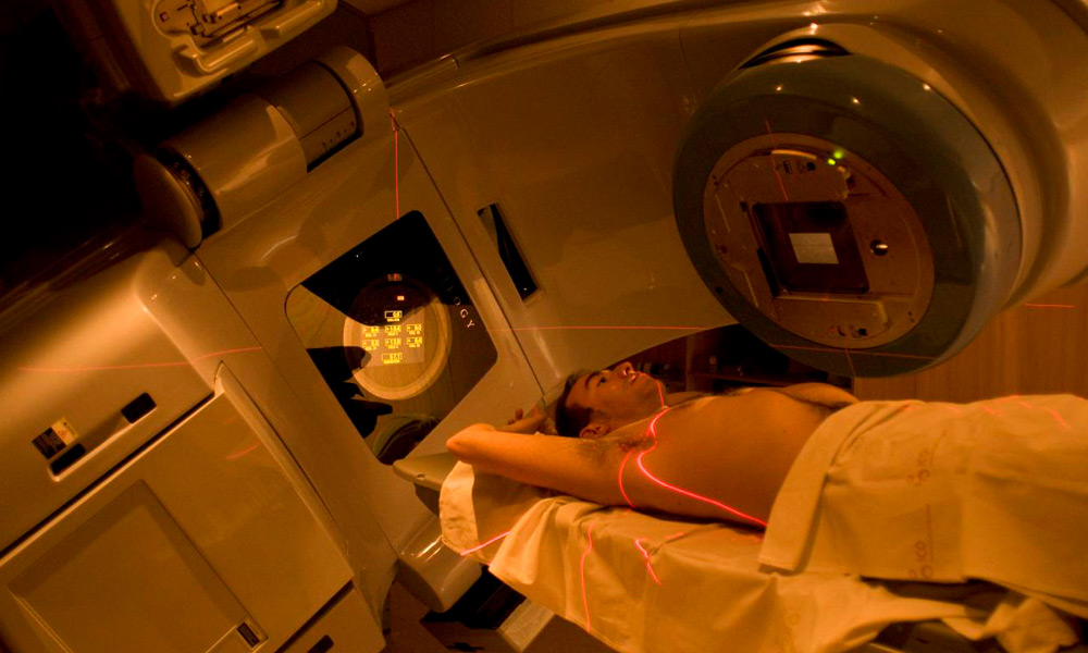
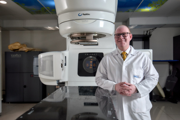
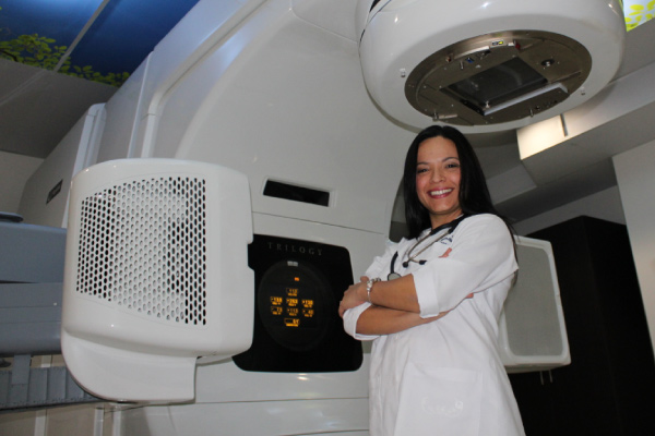
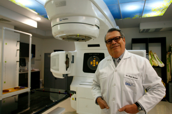
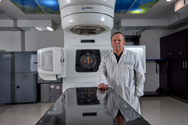
At the Siglo XXI Radiotherapy Medical Center we have extensive experience in the application of the latest technology for the treatment of injuries with radiotherapy. Through the use of IMRT or Intensity Modulated Radiotherapy, patients receive high doses of treatment in a millimetrically precise manner, this translates into a significant decrease in the amount of healthy tissue that is subjected to radiation doses, fewer side effects, better preservation of the healthy tissue and greater optimization of the patient's treatment time (fewer days of treatment than with conventional radiotherapy).

In addition to this, with the new 4D radiotherapy technology, the patient's respiratory cycles are considered in treatment planning. In this way, the equipment and doses will be calibrated to apply the treatment doses in the most optimal phases of the respiratory cycle, increasing the effectiveness and precision of the treatment of mobile tumors or lesions.

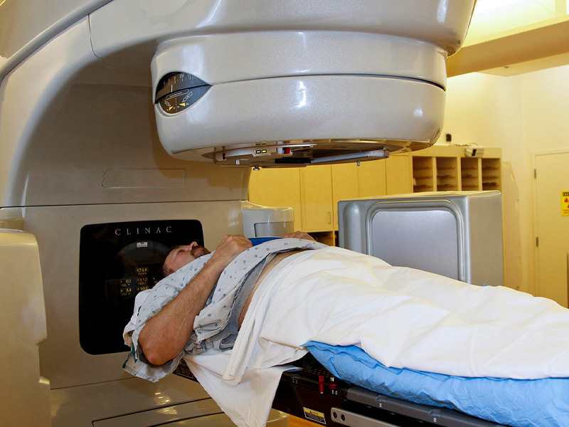
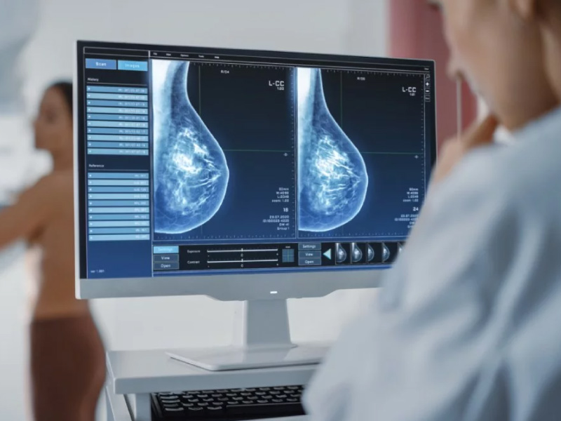
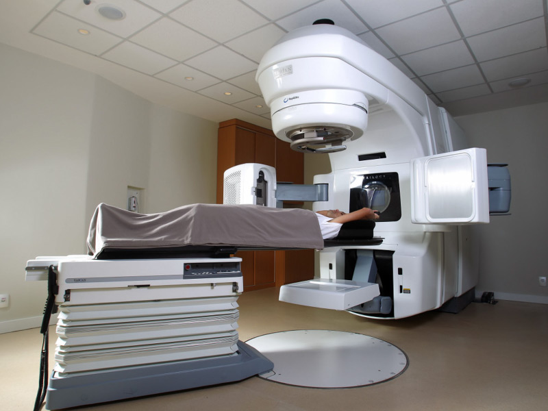
IMAGE GUIDED RADIOTHERAPY (IGRT): Image Guided Radiation Therapy or IGRT combines CT scan images with radiation therapy during each treatment session. This allows the area to be treated to be determined as precisely as possible and the areas surrounding the treatment area to be maximally protected.

What this technology does is place the patient at the exact point to irradiate the injury almost exclusively and not damage important organs; taking into account millimeter variables. This technology is not available in conventional radiotherapy systems.

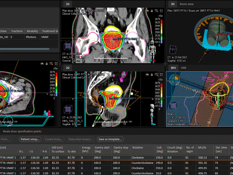
INTENSITY MODULATED RADIOTHERAPY (IMRT): Intensity Modulated Radiotherapy or IMRT is a technology through a computerized system that allows precise doses of radiation to be delivered to a tumor or specific areas within a tumor and reduce doses to healthy tissues.

This technology combined with the advantages of our linear accelerator offer the so-called Arc Therapy, which is the ability to irradiate an injury from 360 different angles, guaranteeing the highest quality treatment in an extremely short time (approximately 3 minutes). In this way, patient comfort and levels of precision are increased.

RADIOSURGERY: High doses in few treatments (1-5 sessions). Radiosurgery or SBRT is used in the treatment of small to medium-sized tumors.
Despite its name and compared to traditional radiation therapy, it is a non-surgical procedure that delivers highly focused radiation at much higher doses and in only one or a few treatments. This treatment is possible thanks to the development of highly advanced radiation technologies that allow the delivery of maximum doses within the target, while minimizing the dose received by the surrounding healthy tissue. The goal is to administer doses that destroy the tumor and achieve maximum local control.

Prostate cancer

Prostate cancer

What is prostate cancer?

Prostate cancer is the second most common type of cancer among men in the United States, and according to the incidence in Costa Rica between the period from 2000 to 2015, prostate cancer was also the most common in men, followed by skin, stomach, colon and lung.

The prostate is a gland that only men have, therefore it belongs to the male reproductive system, it is located just below the bladder and in front of the rectum. Prostate cancer occurs when prostate cells begin to grow out of control.

HOW CAN PROSTATE CANCER BE DIAGNOSE?

The most common tests for prostate cancer are:

RECTAL TOUCH: It consists of an examination in which the doctor puts on a lubricated glove and rectally palpates the prostate through the rectal wall in search of nodules or abnormal areas. The prostate gland is located immediately in front of the rectum, and most cancers begin in the back of the gland, which can be felt during a rectal exam. This test is uncomfortable, but it is not painful and takes very little time to perform.

PROSTATE ANTIGEN TEST: Measures the level of Prostate Specific Antigen, a protein found in a man's blood, produced by the prostate gland. The chance of having prostate cancer increases as the PSA level rises. Most men without prostate cancer have PSA levels less than 4 ng/mL of blood. When prostate cancer forms, the PSA level often rises above 4. However, a level below 4 ng/ml does NOT guarantee that a man will not have cancer. About 15% of men who have a PSA less than 4 will develop prostate cancer if a biopsy is done.

PROSTATE BIOPSY: In which the doctor obtains tissue from the gland to determine if cancer cells are present. For some men, having a prostate biopsy may be the best option, especially if the initial PSA level is high. This test is the only way to know for sure if a man has prostate cancer. If prostate cancer is found in a biopsy, this test can also help indicate how likely the cancer is to grow and spread quickly.

If prostate cancer is found in the biopsy, a grade will be assigned. The grade of the cancer is based on how abnormal the cancer looks under a microscope. This test will confirm the diagnosis of cancer and will give us an idea of the tumor volume and the degree of aggressiveness. The Gleason score, which has been in use for many years, assigns grades when comparing cancer with normal prostate tissue:

SYMPTOMS: 

Need to urinate often, especially at night

Difficulty starting or stopping urine stream

A weak or interrupted passage of urine

Difficulty urinating

Dripping urine when laughing or coughing

Pain or burning when urinating

Blood in urine or semen

A dull pain or stiffness in your lower back, ribs, or upper thighs

Swelling, weakness or numbness of the lower extremities (areola) or the skin of the breast, Redness or small holes in the skin over your breast, like the skin of an orange.

TREATMENT

Among the techniques that are used and provided in 21st Century Radiotherapy are:

INTENSITY MODULATED RADIOTHERAPY (IMRT): Allows the RT dose and intensity to be varied (escalated) during therapy. Reduces side effects and facilitates treatment when pelvic lymph nodes must be included in the field. It allows very high doses (81 Gy) to be administered with little intestinal toxicity. This technique uses a computer-controlled machine that moves around the patient as it delivers radiation. In addition to configuring the beams and directing them at the prostate from various angles, the intensity (strength) of the beams can be adjusted to limit the radiation doses reaching adjacent normal tissues. This allows doctors to deliver an even higher dose of radiation to the cancer.

STEREOTAXIC RADIOTHERAPY (SBRT): It consists of administering a high dose of irradiation to the prostate using complex and precise radiotherapy techniques. Currently, early stages can be treated with this radiotherapy technique in just 5 days in our center with greater precision without increasing the side effects of the treatment.

IMAGE GUIDED RADIOTHERAPY (IGRT): It also allows the administration of higher doses of external radiation therapy (even reaching up to 90Gy) through the use of advanced imaging techniques such as magnetic resonance imaging with spectroscopy or an integrated imaging scanner. This advancement allows the doctor to take photographs of the prostate just before administering radiation to make minor adjustments to the direction of the rays. This appears to help deliver the radiation even more precisely, leading to fewer side effects, and is a novel technique currently being applied at our medical center.

ACTIVE SURVEILLANCE: For very low risk and low risk stages. If you and your doctor agree that active surveillance is a good idea, your treatment will include regular doctor visits and blood tests to measure prostate-specific antigen (PSA). If the cancer grows or your condition changes, your doctor may start treatment
If the cancer has spread beyond the prostate, doctors use systematic treatments. These include hormone therapy, chemotherapy, and vaccines for prostate cancer.

Breast cancer

Breast Cancer and its Treatment

What is breast cancer?

EPIDEMIOLOGY

Breast cancer is the leading cause of death in women in developing countries and the second cause of death in developed countries, second only to lung cancer. Compared with international data, an incidence of 30-39.9 cases/100 thousand inhabitants is documented in Costa Rica and in Latin Americans 4.1, this being lower in relation to the incidence of cases in countries in North America, Australia and North-Western Europe where the incidence is around 78-90/100 thousand inhabitants.

RISK FACTOR'S

Factors that may affect breast cancer risk include:

RISK FACTOR'S: Factors that may affect breast cancer risk include:

AGE:The most important risk factor for breast cancer is age. Most breast cancers occur in women > 50.

FAMILY BACKGROUND: Having a first-degree relative (mother, sister, daughter) with breast cancer doubles or triples the risk of developing it, but the risk in more distant relatives increases only slightly.

GENE MUTATION FOR BREAST CANCER: About 5 to 10% of women with breast cancer carry a mutation in 1 of 2 known breast cancer genes, BRCA1 or BRCA2. If these women's relatives also carry the mutation, they have a 50 to 85% lifetime risk of developing breast cancer.

PERSONAL HISTORY: Having had invasive breast cancer increases your risk. The risk of developing cancer in the contralateral breast after mastectomy is 0.5 to 1%/year of follow-up.

GYNECOLOGICAL HISTORY: Early menarche, late menopause, or a late first pregnancy increase the risk.

BREAST CHANGES: A history of an injury that required a biopsy slightly increases the risk. Benign lesions that may slightly increase the risk of invasive breast cancer are complex fibroadenoma, moderate or florid hyperplasia (without atypia), sclerosing adenosis, and papilloma. An increase in breast density on a screening mammogram is associated with an increased risk of breast cancer.

USE OF ORAL CONTRACEPTIVES: Oral contraceptives increase the risk very slightly (about 5 more cases per 100,000 women). The risk increases primarily during the years of contraceptive use and decreases slowly during the 10 years following discontinuation.

Early detection

Breast cancer is a major health problem worldwide. Despite diagnostic and therapeutic advances, its prognosis continues to depend mainly on the extent of the disease at the time of detection. Hence, getting an early diagnosis remains the best way to improve your chances of cure.

Mammography has been proven to be the most effective diagnostic test; it reduces mortality from breast cancer.

SYMPTOMS: A lump or thickening in the breast that feels different from the tissue around it. A palpable nodule in the breast is the most common sign, generally painless. Change in size, shape or appearance of a breast. Changes in the skin over the breast, such as dimpling.

Recent nipple inversion scaly, peeling, crusting, and peeling of the pigmented area of skin surrounding the nipple (areola) or breast skin, Redness or small holes in the skin over your breast , like the peel of an orange.

DIAGNOSIS: When there is a suspicion of breast cancer either through physical examination (both by the patient and a doctor) or by a routine mammogram, a study is initiated to confirm or rule out that suspicion. Imaging tests will guide the diagnosis, but a certain diagnosis of breast cancer always requires confirmation with a biopsy.

WITHIN THE TESTS ARE

SELF-EXPLORATION: It has not been proven effective in reducing the mortality of the disease.

MAMMOGRAPHS: They are X-ray images that detect abnormal areas in the breast. They do not have a 100% reliability so they may give suspicious images that are ultimately not malignant (false positives) or fail to diagnose a malignant tumor (false negatives).

ULTRASOUNDS: Technique that uses ultrasound to produce an image and can distinguish cystic lesions (fluid-filled, normally non-tumorous) from solid lesions (more suspicious). Many times this technique complements mammography. Ultrasound can also assess the status of the lymph nodes in the armpit, which are the first site of spread of breast cancer.

NUCLEAR MAGNETIC RESONANCE: It is a radiological examination that uses the action of an electromagnetic field to obtain images. It may be necessary in women with dense breast tissue, women with a BRCA gene mutation or women wearing silicone prostheses.

If breast cancer is suspected, the next step is to take a tissue sample for analysis.

BIOPSY: It consists of extracting a sample of tissue from the suspicious area to analyze it microscopically and determine its benign or malignant characteristics, as well as the type of tumor cells, their degree of aggressiveness and some other parameter of interest. when making decisions about treatment.

The biopsy can be done by direct palpation or guided by ultrasound. This can be done with a fine needle (FNAB) or with a thick needle (CNA) to obtain a greater amount of tissue. Sometimes a biopsy may be necessary in the operating room.


TREATMENT

Your doctor determines breast cancer treatment options based on the type of breast cancer, its stage, grade, size, and whether the cancer cells are sensitive to hormones. Treatment will depend on the stage at the time of diagnosis, whether it is ER/PR positive or not, HER2 positive or not. Surgery (stage I, II, IIIA to IIIC), is a pillar of breast cancer treatment. There are two (2) options:

The Sentinel node, the node where the nodal metastasis from the primary tumor is first discovered, has come to replace routine axillary nodal dissection, thereby reducing the morbidity that this entails; such as edema of the upper limb, neuropathies. Sentinel lymph node biopsy is a standard procedure and cannot be replaced by imaging studies such as MRI or CT scan.

Radiotherapy: Used post-surgery in breast conservation (since recurrence is >20%) or in cancer with high risk of local recurrence. Its purpose is to eradicate residual disease. Radiotherapy after breast-conserving surgery significantly reduces the incidence of local recurrences in the breast and regional nodes and may improve the overall survival rate.

The Siglo XXI Radiotherapy Medical Center is the only center in the country that offers high-precision treatments that allow partial or high-dose regimens to be applied that better preserve healthy tissue and in a shorter amount of time.

Below are the details of some of these treatments:

RADIATION TO THE WHOLE BREAST: The conventional schedule for delivering radiation to the whole breast is 5 days a week (Monday through Friday) for 6 to 7 weeks. Another option is hypofractionated radiation therapy in which radiation is also given to the entire breast, but in higher daily doses (Monday through Friday) using fewer treatments (usually for only 3 to 4 weeks).

PARTIAL BREAST IRRADIATION: Intensity-modulated radiation therapy (IMRT): IMRT delivers powerful doses to certain parts of the tumor bed and helps reduce damage to adjacent normal body tissues.

4D RADIOTHERAPY: It is a technique that takes into account the movement of the tumor not only during treatment but also during planning. To do this, the patient's image acquisition system not only takes the images at a given moment, but a series of images are obtained with the treatment area with the different positions it can adopt. It takes into account the patient's respiratory cycles which are considered in treatment planning, in this way the equipment and the dose are calibrated to apply the treatment doses in the most optimal phases of the respiratory cycle.

IMAGE GUIDED RADIOTHERAPY (IGRT): This technique uses high-resolution three-dimensional images to locate the precise location of tumors, adjust the patient's position for maximum precision, and deliver radiation therapy. This allows the area to be treated to be determined in the most precise way and the areas surrounding the treatment area to be maximally protected. This technology places the patient at the exact point that should be treated to almost exclusively irradiate the injury and not damage important organs such as the heart or lungs; technology that is not available in conventional radiotherapy systems.

RADIOSURGERY: High doses in few treatments (5 sessions). Despite its name and compared to traditional radiation therapy, it is a non-surgical procedure that delivers highly focused radiation at much higher doses and in only a few treatments in the case of breast cancer. The adjuvant treatment of breast cancer with external radiation therapy can currently be applied in 5 consecutive days based on the results of the phase III FAST-Forward study (Murray Brunt, 2020) and the Siglo XXI Radiotherapy Medical Center is the pioneer in apply this technique in Costa Rica.

Dr. Rolando Loría

Dr. Rolando Loría

Radiation Oncologist

Founding member of the Costa Rican Radiation Oncology Association. National reference in radiotherapy treatments, being the specialist who treated the most cases in Costa Rica in the period 2022-2023. Extensive experience in treatments of multiple metastases, gynecological tumors and body radiosurgery. 

Academic education

Bachelor of Medicine

Univ. Medical Sciences | Since 2002

Collegiate

College of Physicians and Surgeons CR | Since 2007

Special. Radiation Oncology

University of Costa Rica | Since 2012

Master Advanced Technological Applications in Radiation Oncology

University of Murcia (Spain) | Since 2012

Work experience

EBAIS Coordinator

Health Area in Siquirres|

General Physician in Emergencies

Health Area in Siquirres
| 2006 - 2007

General Physician in Emergencies

Saint Vincent de Paul Hospital | 2007 - 2008

Company Doctor

National Rehabilitation Center | 2007

Resident Doctor Radiotherapy

San Juan de Dios Hospital | 2008 - 2012

Assistant Physician Specialist in Radiation Oncology

Mexico Hospital | 2012 - 2012

Assistant Physician Specialist in Radiation Oncology

Hospital Clínica Bíblica / 2012

Air Ambulance Doctor

AirEvac International / 2014

Postgraduate Professor of Radiation Oncology

University of Costa Rica / 2014

College professor. Health Technologies

University of Costa Rica / 2014

Dr. María Bonilla

Dr. María Bonilla

Oncologist and Radiation Therapist

Specialist with the longest experience in the country in the treatment of head and neck tumors and Oncohematological diseases. Extensive experience in radiosurgery and body radiotherapy treatments, treatments of lymphomas, sarcomas, multiple metastases, among others.

Academic education

Specialty in Radiation Oncology

University of Costa Rica| From 2010

Bachelor's and Doctorate in General Medicine and Surgery

International University of the Americas | Since 2002

Work experience

Assistant physician specialist in radiation oncology

Mexico Hospital | 2011 - Current

3CD and IMRT RT techniques

Madrid Institute of Oncology (IMO) |

General Assistant Physician

San Vicente de Paul Heredia Hospital | 2007 - 2011

General Assistant Physician

Alfaro Ruiz Health Area (EBAIS) | 2003

Dr. Hugo Recinos

Dr. Hugo Recinos

Radiation Oncologist

More than 35 years of experience in Radiation Oncology. Dr Recinos has specialized in brain and body radiosurgery treatments. He has extensive experience treating brain injuries, metastatic tumors, prostate cancer treatments and more.

Academic education

Doctorate in medicine

Autonomous University of Central America, Costa Rica | Since 1984

Internal Medicine Residency

University of Costa Rica | Since 1986

Post – Degree in Radiation Oncology

University of Costa Rica | Since 1989

3D radiotherapy treatment planning

University Hospital of Caracas | Since 1992

Training course in High Dose Rate Brachytherapy for Gynecological Cancer

INCan - Mexico | Since 1994

Training program in Radiation oncology

Comprehensive Cancer Center – Florida | Since 1998

Work experience

Head of the Radiotherapy Service Hemato-Oncology Dept.

San Juan de Dios Hospital
| 2003 - 2016

Assistant Physician in the Radiotherapy Service

San Juan de Dios Hospital.
| 1992 - 2003

Assistant in Internal Medicine. Emergencies

San Juan Dios Hospital
| 1998 - 1994

Social Medical Service

Dr. Carlos Luis Valverde Vega Hospital
| 1986 - 1985

Dr. Luis García

Dr. Luis García

Radiation Oncologist
Medical director

Medical Director and founding member. Dr García has more than 35 years of experience in Radiation Oncology, being one of the doctors who contributed to the improvement of technology for radiation treatments in the country.

Academic education

Doctor of General Medicine

University of Costa Rica | Since 1980

Radiation Oncologist

University of Costa Rica | Since 1986

Post-Graduate Radiotherapy

INCan - Mexico | Since 1987

Post-Graduate Intraoperative Radiotherapy and Hypertemia

National Cancer Center Japan - Japan | Since 1987

High dose rate brachytherapy

Paris - France | Since 2000

Work experience

Director of Postgraduate in Radiotherapy

University of Costa Rica | 1998 - 2005

Radiotherapy Service Assistant

Hospital Mexico - Costa Rica | 1986 - 2005

President of the Scientific Ethics Committee

CIMA Hospital | 2000 - 2010

Medical director

Irazú Radiotherapy Medical Center | 2000 - 2010

Vice president

National Institute of Oncological Clinical Studies | 2000 - 2010

President

CIMED Medical Imaging Center | 2012 - 2016

President

Nuclear Medicine - Cima Hospital

Understanding the importance of adequate time between each radiotherapy session

Understanding the importance of adequate time between each radiotherapy session (rests and exposure to the radiation beam)

By: Dr Rolando Loría

Radiotherapy It is a treatment commonly used in the management of various types of cancer. Radiation is given in fractions (sessions) to maximize damage to cancer cells while minimizing the impact on surrounding normal tissues. The rate of cell death is a crucial aspect in the effectiveness of radiotherapy treatment.

When we talk about the medical term “intra-fraction elongated intervals,” we are referring to the time separation between radiation doses within a treatment. Administration of fractionated doses allows normal cells time to repair damage caused by radiation, while cancer cells, which often have a more limited repair capacity, can accumulate damage and eventually die (Shibamoto, Miyakawa, Otsuka , & Iwata, 2016).

If the times between doses are too short, normal cells may not have enough time to recover, which may increase the risk of side effects. On the other hand, if the intervals are too long, cancer cells may have time to repair some of the damage, reducing the effectiveness of the treatment.

The average times between each radiotherapy session They will depend on the dose and the treatment scheme, however the minimum “rest” time between one session and another is approximately 6 hours in Brain injuries and 8 hours in body injuries.

Maximum “rest” times They will vary between 24 or 48 hours between each session depending on the indicated treatment scheme.

EXHIBITHION TIME

In addition to this, there is another important variable to consider: the times of exposure to radiation during treatments.

The expert Khorramizadeh and collaborators demonstrated that when the exposure time to the radiation beam per session increases from 15 minutes to 45 minutes in each fraction (treatment session), the death of tumor cells is reduced. It seems that the main phenomenon that affects the cellular response is the repair of sublethal damage. The effect of the dose administered over 15 minutes may be greater than that of 30 and 45 minutes (Khorramizadeh, Saberi, & Tahmasebi-Bi, 2017).

High dose equipment such as the Linear Accelerator that the XXI Century Radiotherapy Medical Center, They allow radiosurgery doses to be delivered extremely accurately in the maximum periods indicated in these studies, achieving not only the expected cellular damage but also contributing to the patient's comfort by not being exposed to long periods of treatment as happens in other radiosurgery equipment.

The rate of cell death in radiotherapy depends on several factors, including the sensitivity of the cells to radiation damage, the type of radiation used, the total dose administered, and the dose fraction. The relationship between cell death rate and specific intra-fraction intervals may vary depending on the type of cancer and treatment (Benedict, Lin, Zwicker, Huang, & Schmidt-Ullrich, 1997).

It is important to note that radiotherapy treatment planning is carried out in a personalized manner for each patient, taking into account various clinical and radiobiological factors. Radiation oncologists work to optimize the delivery of radiation to achieve maximum effectiveness against cancer while minimizing side effects on normal tissues.

Bibliography

Khorramizadeh, M., Saberi, A., & Tahmasebi-Bi, M. (September 1, 2017). Impact of Prolonged Fraction Delivery Time Modeling Stereotactic Body Radiation Therapy with High Dose Hypofractionation on the Killing of Cultured ACHN Renal Cell Carcinoma Cell Line. J Biomed Phys Eng(3), 205-216.

Shibamoto, Y., Miyakawa, A., Otsuka, S., & Iwata, H. (2016). Radiobiology of hypofractionated stereotactic radiotherapy: what are the optimal fractionation schedules? J Radiat Res.(57 Suppl 1), 76-i82.

Benedict, S., Lin, P., Zwicker, R., Huang, D., & Schmidt-Ullrich, R. (Mar 1, 1997). The effectiveness of intermittent biological irradiation as a function of overall treatment time: development of correction factors for linac-based stereotactic radiotherapy. Int J Radiat Oncol Biol Phys., 37(4), 765-9.